
Scientists have developed a new visual express method of searching for diseases
0
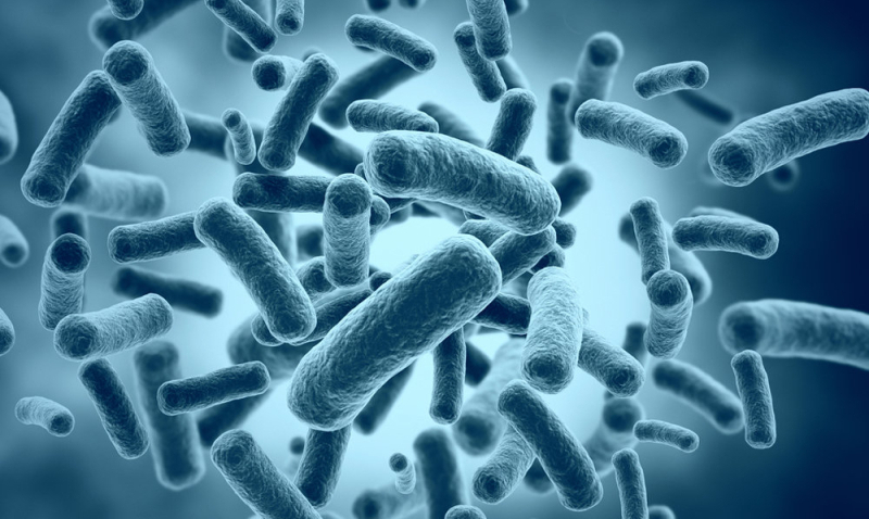
American scientists have developed a new method of fluorescent bioimaging, which allows real-time observation of processes occurring in the body, search for foci of diseases and control of treatment. The description is given in the journal Nature Communications.
Details
The effectiveness of molecular imaging using fluorescent agents – fluorescent bioimaging – has been proven at the level of laboratory and clinical trials. In particular, this method is used to control the removal of tumors during oncological operations on the lungs, as well as during pre- and intraoperative observation of patients. Various variants of the method are widely used by biologists and doctors to study various processes occurring in living tissues of animals and humans.
The essence of the method is that a contrasting fluorescent agent is introduced into the tissue or blood, which has the ability to bind to specific cells (for example, cancer cells) or proteins. When tissues are exposed to light of a certain spectrum zone, these cells become visible.
However, the fluorescent bioimaging method has a drawback — due to the complexity of biological data collection and processing systems to construct an image, there is a need for labor-intensive calculations that take a lot of time.
American scientists from the University of Southern California, in collaboration with colleagues from the University of Cambridge, have developed a new a variant of the molecular imaging method that can work in real time.
As a basis, they took the technique of fluorescent hyperspectral visualization, which allows not only to monitor the observed molecules, but also to differentiate their colors over the entire spectrum, as well as to create full-color images of the internal parts of the body. The authors' modification of this technique, called the Spectrally Encoded Enhanced Representation (SEER) method, provides 2.7 times
greater image clarity than existing methods and is 67 times faster.
The speed is ensured by a special algorithm for processing the signals of fluorescent labels, and a higher level of detail is achieved due to the analysis of signals across the entire spectrum. But most importantly, this method uses less computer memory, which is very important for large-scale research.
“SEER is a fast and intuitive mathematical way to interpret images as they are collected and processed. This is a kind of element of basic science that can be used for the development of better methods of diagnosis and treatment – the words of the head of the study Francesco Cutrale, professor of molecular and computational biology at the University of Southern California, are quoted in the press release of the university. – This technology is quite universal. It can become the basis of a smartphone program for use in remote medicine, determination of food safety”.
In the near future, scientists together with doctors at the Children's Hospital of Los Angeles plan to use the SEER method for clinical detection of early stages of lung cancer. as well as to study the effects of exposure to pollutants on the body.


Leave a Reply